
A complete analysis of mammography: a health tool that American women must know
Breast cancer is one of the most common cancers among American women, and early detection and treatment are crucial to improving the cure rate. Mammography (also known as mammography or Mammogram) is the main means of screening for breast cancer and is widely recommended for regular physical examinations. Although this examination is of great significance for the prevention and early diagnosis of breast cancer, many women still have doubts about its process, precautions and possible discomfort. This article will introduce the principles, processes, applicable populations, precautions of mammography in detail, and combine real cases to help American women scientifically understand and actively participate in breast health management.
1. What is a mammogram?
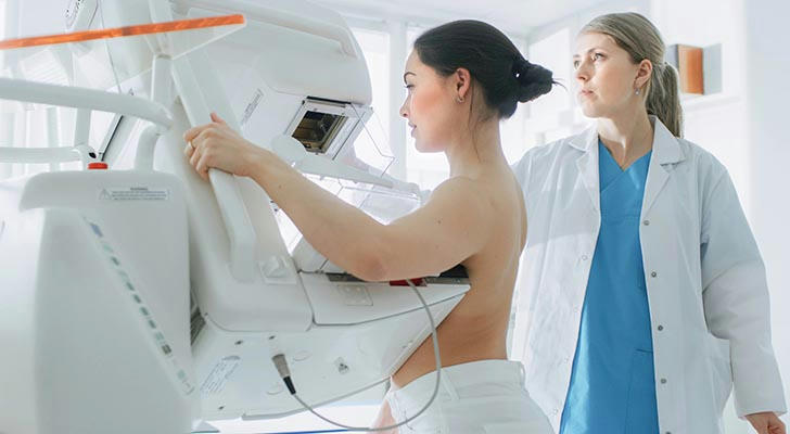
Mammography is a technology that uses low-dose X-rays to image the breast. During the examination, the breast is gently clamped between two plastic plates to flatten the breast tissue to obtain a clear image. Through these images, doctors can find possible lumps, calcifications or other abnormalities in the breast, especially those early cancerous lesions that have not yet been touched.
Although compression of the breast may cause temporary discomfort, it is a necessary step to ensure clear images, reduce radiation doses and avoid image blur. The whole process usually lasts 15 to 30 minutes, and you can resume normal activities after it is completed.
2. Who needs a mammogram?
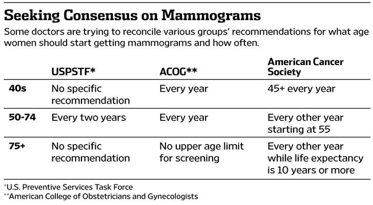
The American Cancer Society recommends that women at average risk of breast cancer should start annual mammograms between the ages of 40 and 45, annually between the ages of 45 and 54, and every two years for those 55 and older. For women with a family history of breast cancer, previous breast disease, or other high-risk factors, doctors may recommend starting screening earlier or more frequent examinations.
In addition, a diagnostic mammogram should be performed promptly when the following symptoms occur:
Unexplained lumps or thickenings in the breast or armpit
Dimples or abnormal changes in the breast skin
Abnormal nipple secretions
Noticeable changes in breast shape or skin color
3. Detailed explanation of the mammogram process
Appointment and preparation
It is recommended to schedule the examination one week after the end of menstruation, when the breasts are less sensitive and discomfort is reduced. Avoid using products such as perfume, deodorant, lotion, etc. on the breasts or underarms on the day of the examination to avoid affecting the image quality. Wear two-piece clothing for easy removal.
Examination process
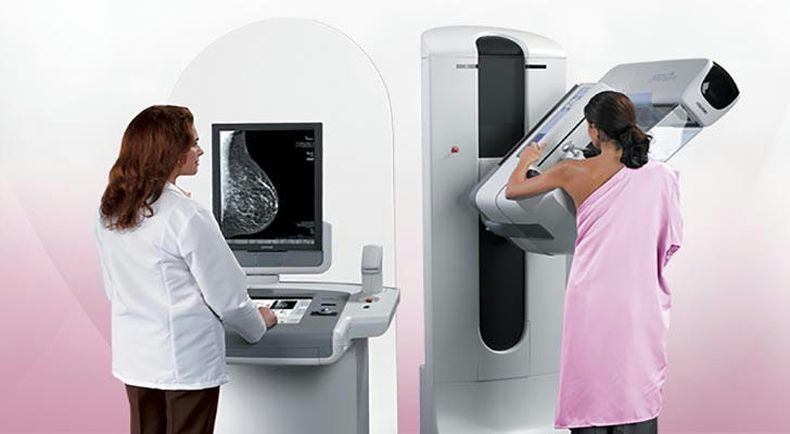
After entering the examination room, the technician will instruct the patient to remove the clothes above the chest and put on the hospital gown. The patient stands in front of the mammogram machine, places one breast on a flat tray, and another transparent plastic plate is slowly pressed down for a few seconds to expand the breast tissue. The technician will adjust the angle and take different views. Then repeat the same steps to check the other breast.
Image inspection and feedback
After the shooting is completed, the technician will check the image quality and may repeat some images if necessary. The entire examination process generally does not exceed 30 minutes. The images are interpreted by professionally trained radiologists, and the report results are usually fed back to the patient and the attending physician within a few days.

4. Precautions and common questions for mammograms
Will it hurt?
There may be a short-term sense of pressure or slight discomfort when pressing the breast, and some women may feel pain. You can inform the technician in advance and adjust the pressure if necessary.
Is radiation safe?
The radiation dose used in mammograms is extremely low, far lower than the natural radiation exposure in daily life, and the risk is extremely small. The benefits of early cancer detection far outweigh the radiation risks.
Do I need to fast before the examination?
No, you can eat and take medicine normally.
Can patients with breast implants be examined?
Yes, but you need to inform the technician and use special technology to take the picture.
How to avoid false positives or false negatives?
Regular examinations, combined with doctor's advice and other imaging examinations (such as ultrasound and MRI) can help improve the accuracy of diagnosis.
5. Sharing of real cases
Case 1: Mary's early discovery
Mary, 52 years old, has a mammogram every year as recommended by the American Cancer Society. During a routine examination, the doctor found tiny calcifications in her breast. Although there was no obvious lump, further biopsy was recommended. It was eventually diagnosed as early breast cancer. After surgery and adjuvant treatment, Mary recovered well. She said that regular mammograms have helped her avoid the risk of advanced cancer.
Case 2: Susan's examination experience
Susan, 45 years old, had her first mammogram. She was worried about the pain of the examination. The technician patiently explained the process and used the MammoPad protective pad to reduce the pressure. The examination was completed smoothly, the image was clear, and no abnormalities were found. Susan said that after understanding the details of the examination, she felt relaxed and recommended regular screening for women around her.
Case 3: Screening strategies for high-risk groups
Linda has a family history of breast cancer and began to undergo mammograms and MRI screening at the age of 40. No abnormalities were found in multiple examinations, but she insisted on annual physical examinations and closely monitored her breast health. The doctor developed a personalized screening plan based on her risk situation and adjusted the frequency and items of examinations in a timely manner.
Conclusion
Mammograms are an important tool for women's breast health management. They can detect breast cancer early and significantly improve the success rate of treatment. Understanding the examination process, arranging time and precautions reasonably can help reduce discomfort and improve the effectiveness of the examination. Real cases have proved that regular mammograms are essential for preventing breast cancer. In the face of breast cancer risks, actively participating in scientific screening is a wise choice for every woman to protect her health. It is recommended to arrange mammograms reasonably according to personal risks and doctor's advice, and work together to build a healthy future.